
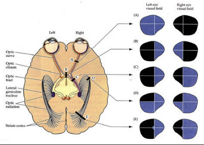
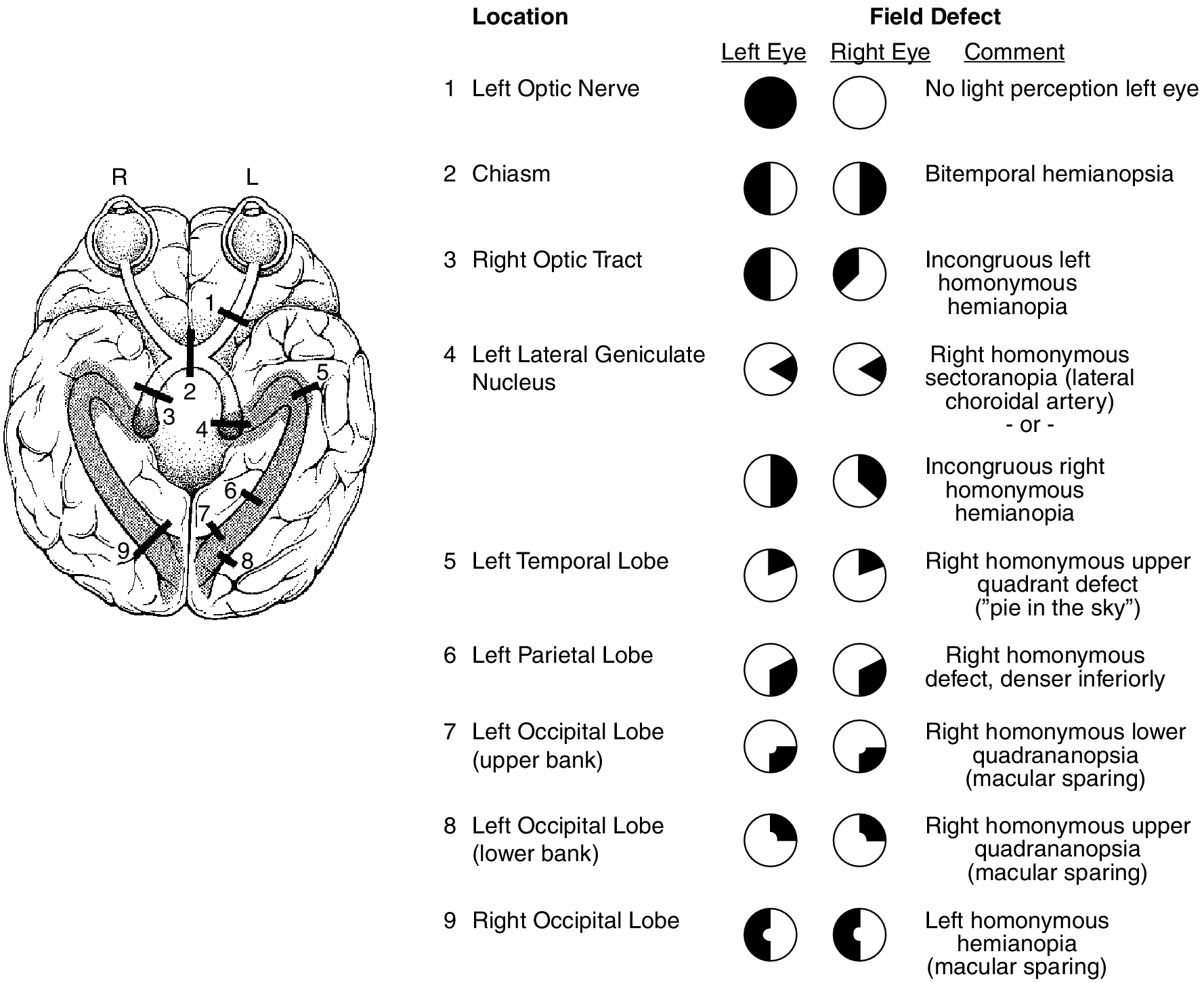
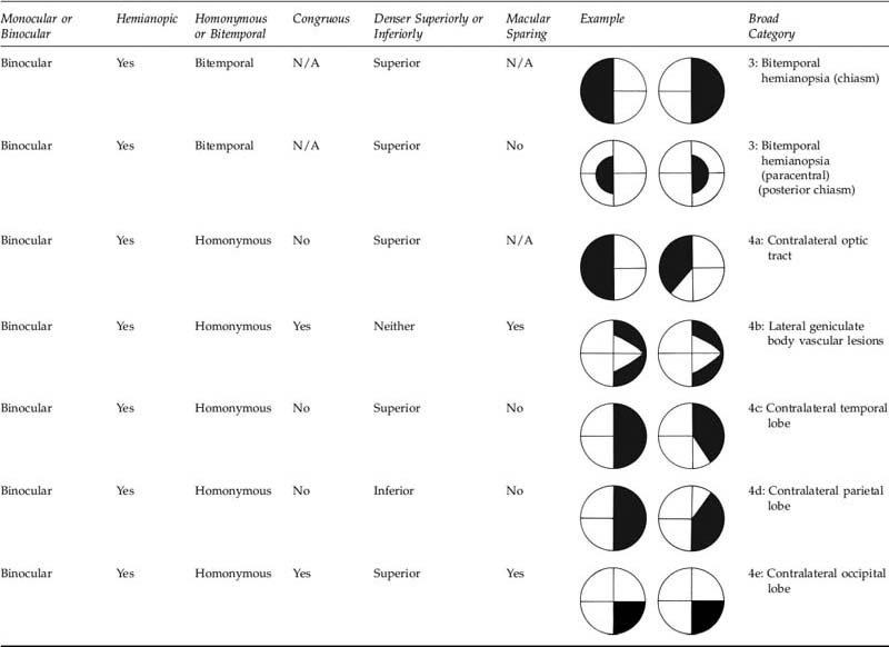
At the optic chiasm fibres from the nasal half of the retina corresponding to the temporal visual field decussate. Lesions to optic radiations result in homonymous contralateral quadrantanopia.

Characteristic Visual Field Defects Of Patients With Occipital Lobe Infarction Homonymous Hemianopia And Macular Sparing Springerlink
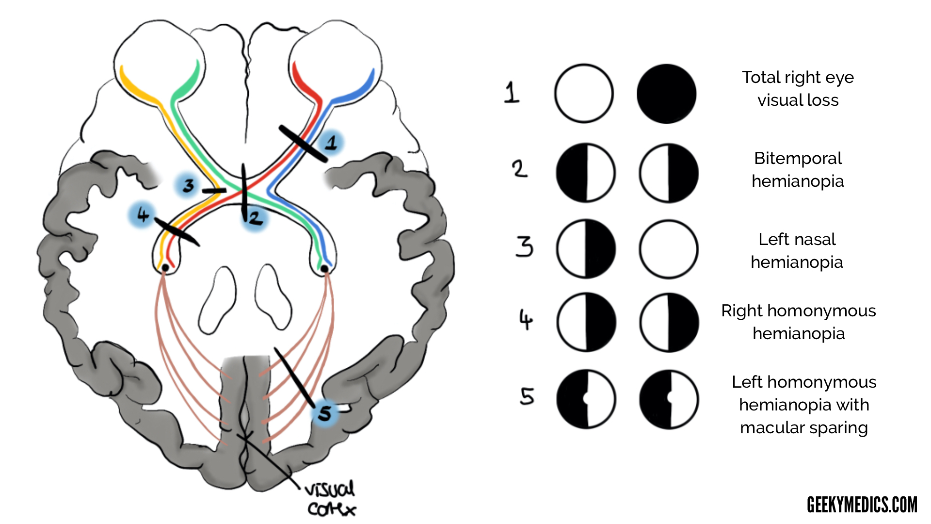
Visual Pathway And Visual Field Defects Geeky Medics
Visual Field Defects On Meducation

Question submitted by Andrew Hendrick MD PGY-2 Ophthalmology Resident at the University of Colorado in Aurora.

Visual field defects. Visual field loss can be diffuse as with cataract or corneal opacification but more commonly there are isolated defects. Bilateral glaucoma bilateral retinopathy retinitis pigmentosa. For information about all other fields see Work item field index.
It states that Computer enhanced perimetry involves the use of a micro-computer to measure visual sensitivity at pre-selected locations in the visual field. The results of the analyser identify the type of vision defect. Glaucoma is a disease characterized by high eye pressure Most types of glaucoma begin with loss of peripheral vision.
Understanding the rehabilitation options available and where to refer. The Examination of the Eye article offers further details. Near-normal and severely damaged visual fields will both have low PSD.
Depending on what the history and findings so far suggest a full neurological examination or further tests on the eye may be warranted. It is a covered service when used in assessing visual fields in patients with glaucoma or other neuro-pathologic defects. The visual pathway consists of structures that carry visual information from the retina to the brainLesions in the pathway cause a variety of visual field defects.
Vigabatrin VGB is an antiepileptic medication used in the treatment of infantile spasms IS and refractory complex partial seizures. During binocular viewing the fields of the two eyes substantially overlap. And the points selected cover the areas that are known to be susceptible to glaucomatous defects.
The gold standard in visual field testing Humphrey Field Analyzer 3 now combines everything you value in a Humphrey with expanded testing options and reduced patient test times. Visual field defects may stem from neurological or ophthalmic problems. The most common visual field test uses a light spot that is repeatedly presented in different areas of your peripheral vision.
Visual rehabilitation aims to maximise the residual vision and decrease functional disability. The same holds true for binocular defects that occupy different positions in space. Find Out More about Heru for Clinicians.
The visual field test is a subjective measure of central and peripheral vision or side vision and is used by your doctor to diagnose determine the severity of and monitor your glaucoma. The patient is asked to look at the dot one eye at a time and note whether the grid lines surrounding the dot appear distorted faded or partially missing. Results Available in Real-Time.
Patient Takes Visual Field Test. A central scotoma involves only fixation whereas a ceco-centralscotomainvolves fixation. A visual field test is commonly used to diagnose or monitor glaucoma.
This is a common test to evaluate for scotomas and also evaluate any visual field restrictions from eyelid disorders like ptosis droopy upper eyelid. The magnitude of the deepest defect across all meridians as well as the difference in. Visual fields with the age-normal sensitivity at each point will have a PSD of 0 as will visual fields in which each point is uniformly depressed from the age-normal value.
For information about fields specific to the CMMI process Bug see Bugs issues and risks field reference. 5252018 3 Factors influencing visual field measurements RESPONSE FACTORS Patient instructions Patients expectations Examiners personality Response criterion The patients willingness to say yes when a target is presented Strict criterion higher threshold Relaxed criterion lower threshold Reaction time In general the more peripheral the stimulus the longer. Visual field defects after stroke A practical guide for GPs Background Visual field defect after stroke can result in significant disability and reduction in quality of life.
This test is most often used to detect central visual field defects. Use the fields described in the following table to capture both the initial issue and ongoing discoveries. Patient uses a wearable device to easily take the visual field test that is smart fast and provides immediate results.
It is important to understand that the image of an object in the visual field is inverted and reversed from left to right much like the image on film from a camera lens. Lesions compressing the chiasm such as pituitary adenomas therefore cause bitemporal hemianopia. Visual field defects.
The type of field defect can help localize where the lesion is located see figure. There is no cure for glaucoma stopping the progression of the disease will help preserve vision. Thus the largest PSD will be registered for focal deep visual field defects.
The visual field exam is a crucial part of glaucoma diagnosis and is repeated periodically to determine if the disease is stable or getting worse. Therefore it provides information regarding the location of any disease processes or lesions throughout the visual. If visual field defects are seen ethambutol should be discontinued and follow-up fields should be obtained every 13 months till visual fields have either improved or stabilized.
This is a printed image of a grid with a dot in the center. Peripheral vision test Humphrey visual field exam and the Goldmann visual field exam are among the other names that you may hear regarding visual field testing. The PD plot is designed to highlight localized defects by removing generalized visual field loss likely due to a cataract.
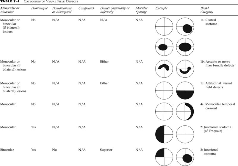
Visual Field Defects Arcuate Central Ceco-central Nasal step Altitudinal Temporal wedge 49 Whats the difference between a central and a ceco-central scotoma. Visual field defects. Phu and Kalloniatis examined the 10-2 and 24-2 test results of 73 patients with central visual field defects.
Visual field tests are a method an eye doctor opthalmologist or optometrist can use to measure how wide an area you can visualize away from a focal point. Perimetry has long been an important tool for identifying visual field defects and guiding glaucoma patients treatment. The Bug work item type uses some bug-specific fields.
Visual field testing should be carried out in each eye separately. Visual Field Testing as a Diagnostic Tool. True defects on the PD should be characterized by their shape and location ie nasal steps central and arcuate scotomas.
Visual defects that result from pathway interruption at any point from the retina to the cortex are described in terms of the visual field rather than the retina. Group 1 Car and motorcycle Group 2 Bus and lorry. For immediate evaluation of vision defects.
Humphrey field analyser HFA is a tool for measuring the human visual field that is commonly used by optometrists orthoptists and ophthalmologists particularly for detecting monocular visual field. The visual field defects associated with glaucoma appear to be fairly non-specific although typical loss fits with the arrangement of the retinal ganglion cell axons within the retinal nerve fibre layer of the retina. Consequently a visual field defect in one eye will still register as having normal vision when both eyes are open.
How To Examine The Visual System Part 1 Visual Acuity Visual Fields And Eye Movements Eye News
H53 413 Visual Field Defect Decision Maker Plus
Neuro Ophthalmology Question Of The Week Emergency Department Evaluation Of Visual Field Defects Neuro Ophthalmology
Visual Field Deficits Chapter 77 Neurologic Differential Diagnosis

Patterns Of Binocular Visual Field Loss Derived From Large Scale Patient Data From Glaucoma Clinics Ophthalmology

Visual Field Defects After Uneventful Vitrectomy For Epiretinal Membrane With Indocyanine Green Assisted Internal Limiting Membrane Peeling American Journal Of Ophthalmology
Visual Field Defects Ento Key
Visual Field Defects Ento Key


